
Ces Urol X:X | DOI: 10.48095/cccu2026005
Robot-assisted nephroureterectomy (RA-NUE) – video
- 1 Urologická klinika LF UK a FN Plzeň
- 2 Klinika zobrazovacích metod LF UK a FN Plzeň
- 3 Šiklův ústav patologie, LF UK a FN Plzeň
- 4 Onkologická a radioterapeutická klinika LF UK a FN Plzeň
Introduction: The indications for nephroureterectomy (NUE) for upper tract urothelial carcinoma (UTUC) are relatively clear, but questions remain regarding the technique used to perform NUE and the indications for lymphadenectomy (LND – lymph node dissection) during NUE. There is a wide range of NUE techniques (open surgery, laparoscopy, endoscopy). Basically, there is a nephrectomy phase and an ureterectomy phase, and possibly also LND, with individual techniques being combined. Open NUE remains the dominant technique for advanced tumors, otherwise minimally invasive techniques are widely used. After many years of gradually performing three methods of laparoscopic NUE, we were able to switch to the complete minimally invasive RA-NUE technique with the purchase of the da Vinci Xi robotic system. No change of position, no need for endoscopy. We present our technique in a video.
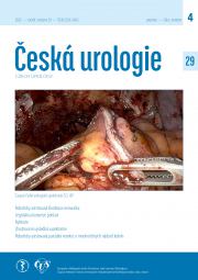
Method: From July 2021 to December 2025, we performed NUE in 93 (85.3%) of 109 UTUC cases and kidney-sparing surgery in 16 (14.7%). RA-NUE from NUE in 43 (46.2%) selected patients, 5 of whom had LND (high-risk cT2–3 or high-grade and cN0). RA-NUE was performed twice for a final histology other than UC – clear cell renal carcinoma and xanthogranulomatous pyelonephritis. In another 5 (4.6%) patients, RA-NUE was performed followed by RA radical cystectomy. Laparoscopic NUE was performed in 11 (10.1%) cases when the robotic system was unavailable. Open surgery (34, 31.2%) was indicated for patients with advanced findings, who were significantly obese, or who had undergone previous extensive intraabdominal surgery. The surgical technique presented in the video: RA-NUE on the left with upper lymphadenectomy: A 4-arm da Vinci Xi system, urinary catheter, and 60° lateral position are used. The most caudal port is 12 mm for stapler application, the others are 8 mm. The assistant port is more medial, 15 mm for final insertion of the Endocatch™ bag II extraction bag. Camera 30°, ProGrasp™, Maryland™ bipolar grasper, monopolar scissors, or SynchroSeal™ sealing tool. The procedure begins with a standard nephrectomy, but the ureter is clipped early to minimize UC dissemination into the bladder. This is followed by paraaortic or paracaval LND using scissors or a sealing device. Without re-docking the system, the ureter is excised craniocaudally in this direction up to the pelvis. The testicular vein is usually preserved, and in women, the ovarian vein is preserved and the entire adnexa is folded medially. The video shows a woman after a panhysterectomy. The ureter is excised from the bladder with the bladder cuff and the bladder defect is sutured with V-Loc® 90 3-0. The suture is applied to the bladder before complete transection of the ureter, which is used for traction. The upper LND can also be performed at the end of the procedure. Extraction of the urinary catheter on the 6th postoperative day after elective cystoradiography (CRG). We do not use intravesical single-dose cytostatics.
Results: A total of 45 RA-NUE in 29 men and 16 women. Average age 71.4±9.3 (47.7-86.1) years. Body mass index 28.0 ± 3.77 (20.2-35.7) kg/sqm. In 38 cases, the described descending technique was used, 7 times with redocking to the pelvic part in the Trendelenburg position. Of these 7 cases, 2 were for distal ureteral tumors started in the pelvic phase, 1 was excised with a bladder diverticulum, and 4 were forced to change to the Trendelenburg position in the second phase (twice for larger distal ureteral tumors, once for a ruptured ureter juxtavesically and intramural part excised by TUR, and once for suspected UTUC based on RA nephrectomy preparation and supplemented by ureterectomy). The average duration of surgery for all 153±29 (99–240) min – for descending RA-NUE 143±21 (99–199) min, with LND 189±35 (164–240) min. SynchroSeal™ was used in the last 17 procedures. Blood loss was 55.8 (0–400) mL. LND was performed in 5 patients (4 women, 1 man) – 4 upper LNDs for pelvic tumors, 1 pelvic LND for a distal ureter tumor – here, the aforementioned change to the supine position was performed, along with excision of the distal ureter and LND. Average number of nodes was 6 (2–10). Hilum renal vessels interrupted by robotic stapler 25× en bloc and 2× separately, 1× separately by external stapler and 17× separately by lockable clips by assistant. Drain used 15×. The weight of the specimen was 496 (128–1,045) g. Histologically, as mentioned, 43× urothelial carcinoma (UC) pTa–pT3 (1× pN1), 1× clear cell renal carcinoma pT2aG3, and 1× xanthogranulomatous pyelonephritis were confirmed. The length of hospitalisation was 7.2 (3–18) days. The catheter was removed on average after 12.8±11.4 (5–58) days (9× left in place for longer than 14 days due to contrast agent leakage during CRG). Complications (except for leakage from the bladder suture) according to Clavien-Dindo – 1× type 2 (prolonged chylous ascites treated conservatively, duration 2 months). Long-term follow-up is not yet available, 1× local recurrence of G3 tumor treated with cystectomy.
Conclusion: RA-NUE has become the standard NUE method at our facility, replacing the laparoscopic approach. So far, it has been optimal for findings where LND is not indicated. The main relative complication is prolonged urinary leakage from the bladder suture. Open NUE remains in the armamentarium for selected cases unsuitable for RA-NUE (advanced tumors, severe obesity, history of extensive intra-abdominal surgery).
Keywords: nephroureterectomy – laparoscopy – robot
Received: January 6, 2026; Revised: February 3, 2026; Accepted: February 11, 2026; Prepublished online: March 3, 2026
video
