


 90 / 100

336
Ces Urol 2016; 20(4): 332–338
KAZUISTIKA
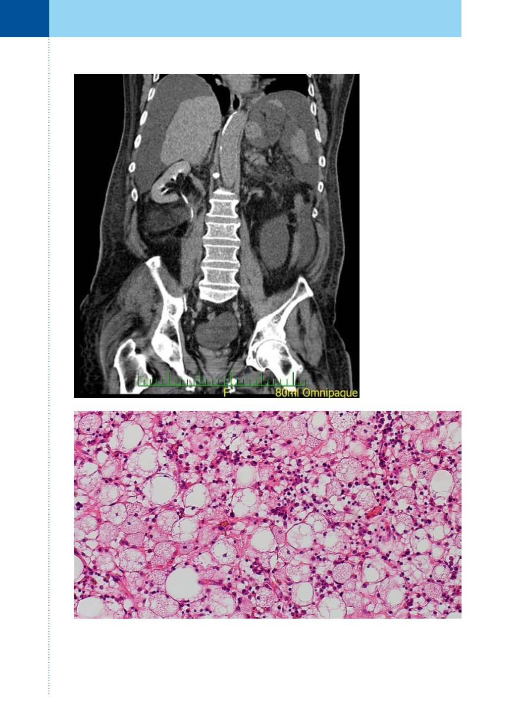
Obr. 5.
Histopatologický nález (hematoxylin-eosin, zvětšení 100×, Nomarského diferenciální interferenční
kontrast, foto: J. Novák)
Fig. 5.
Histopathological finding (H&E stain, magnification 100×, Nomarski differential interference contrast,
photo: J. Novák)
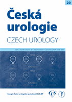
Obr. 4.
CT, koronární
řez, vylučovací fáze: kon-
trolní vyšetření po třech
měsících od exstirpace
tumoru bez průkazu reci-
divy, jako vedl. nález pa-
trný ascites perihepaticky
Fig. 4.
CT scan, coro-
nal section, excretory
phase: control imaging
three months after the
tumour exstirpation with
no signs of relapse; ascitic
fluid surrounding liver is
apparent