


 83 / 100

329
Ces Urol 2016; 20(4): 326–331
KAZUISTIKA
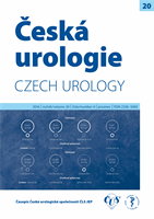
Obr. 3.
Histologické vyšetření preparátu tumoru ledviny: karcinosarkom v levé části obrázku, v pravémhorním
rohu parenchym ledviny s glomeruly, zvětšení 100x
Fig. 3.
Histological specimen of kidney tumor – carcinosarcoma on the left part of the picture, on the right part
renal parenchyma with glomeruli, ZOOM 100x
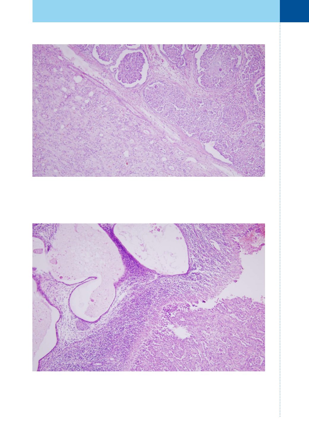
Obr. 4.
Histologické vyšetření preparátu tumoru ledviny: ložisko endometriózy s endometriálními žlázkami,
zvětšení 100x
Fig. 4.
Histological specimen of kidney tumor – focus of endometriosis with endometrial glands on the left part
of the picture, ZOOM 100x